
Endoscopic and Percutaneous Stone Surgery in Abu Dhabi: Ureteroscopy, RIRS and PCNL
When a stone will not pass and lithotripsy is not the right answer, there are two operative routes. Ureteroscopy goes up the natural passage with a fine telescope and breaks the stone with a laser, taking the fragments out rather than leaving them to pass; percutaneous nephrolithotomy makes a track through the flank directly into the kidney, and is what large or staghorn stones need. Both are keyhole operations with no open incision, and which one applies is decided by the size and position of the stone, not by preference.
- Confidential consultation
- Consultant Urologist & Andrologist
- NMC Royal Hospital, Abu Dhabi
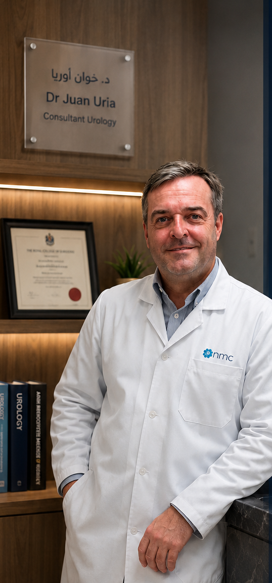
Dr. Juan Uría González-Tova · Consultant Urologist & Andrologist · NMC Royal Hospital Khalifa City, Abu Dhabi
Medically reviewed by Dr. Juan Uría · August 20, 2026
Take the self-assessmentTwo minutes. Nothing is sent or saved.Ureteroscopy covers two related operations. A semi-rigid scope reaches stones in the ureter; a flexible scope goes further, around the bend into the kidney itself, which is what retrograde intrarenal surgery means. In both, a holmium or thulium laser fragments the stone and a basket removes the pieces, so the patient does not have to pass anything afterwards. Stone-free rates are high — considerably higher than lithotripsy for most stones — and the operation is done under general anaesthetic as a day case or a single overnight stay.
Percutaneous nephrolithotomy is the answer to volume. Where a stone is larger than about two centimetres, or fills the collecting system as a staghorn, no amount of laser through a fine scope will clear it in reasonable time. A needle is passed through the skin of the back into the kidney under imaging, the track is dilated, and a larger instrument removes the stone in pieces through it. It is the most invasive of the three stone treatments and the most effective for large stones, needing two to three days in hospital and carrying a real if small risk of bleeding.
The stent is the part patients ask about afterwards, and it is worth explaining before. A ureteric stent is a soft tube left between kidney and bladder to keep the passage open while swelling settles, and it is common after ureteroscopy. It is not painful in the way a stone is, but it does cause urinary frequency, urgency and a dragging discomfort in the flank when passing urine — normal, temporary, and much easier to tolerate when it has been described in advance rather than discovered. It is usually removed within one to two weeks.
Removing the stone is only half the job. Around half of stone formers make another within five to ten years if nothing changes, and the piece of stone that comes out is the single best clue to why. Sending it for analysis, together with blood and 24-hour urine chemistry, identifies a treatable cause in a substantial share of people — and the measures that follow are mostly fluid, salt and diet rather than another operation.
Check your own symptoms
Which stone operation fits your stone?
A structured screen written for this page, built from the stone characteristics that decide the route: size, position, what has already been tried, and what you want weighted. It reads your CT report with you; it does not replace the surgeon reading it.
Have your CT report to hand if you have one. Size in millimetres and where in the kidney or ureter the stone sits are what decide between going up the natural passage and making a track through the flank.
Question 1 of 7
Choose the closest answer
What the assessment involves
Choosing between the two operations — and between them and lithotripsy — is a reading of the CT scan alongside what matters to the patient. Size, position, density and the anatomy of the kidney decide most of it.
A non-contrast CT with the stone measured in three dimensions and its density in Hounsfield units, which together decide the route.
Where in the kidney it sits. A lower-pole stone behaves differently from one in the renal pelvis, and that difference often decides between flexible ureteroscopy and a percutaneous track.
A urine culture cleared before the date, not on it. Operating on an infected system is the one thing that turns a routine stone operation into a serious illness.
Anticoagulants and antiplatelet drugs. These matter far more for a percutaneous track than for a ureteroscopy, and the plan is made with the doctor who prescribed them.
Kidney function and whether both kidneys work, since a single functioning kidney changes both the urgency and the choice of approach.
What you want weighted — the fewest procedures, the shortest recovery, or avoiding a stent. These pull in different directions and are better said out loud than assumed.
Common questions
01Which is better — lithotripsy or ureteroscopy?
Neither, in the abstract. Ureteroscopy clears more stones in one session and takes the fragments away, but it needs a general anaesthetic and often a stent. Lithotripsy needs neither, but leaves you to pass the fragments and more often needs repeating. For a small stone in a favourable position, lithotripsy is a reasonable first choice; for a hard stone, a lower-pole stone, or when being clear after one procedure matters most, ureteroscopy is the better answer.
02How long will I be off work?
After a ureteroscopy, most people are back to desk work within a few days, though a stent in place makes that less comfortable than it sounds and heavy physical work should wait until it is out. After a percutaneous operation, expect two to three days in hospital and around two weeks before normal activity, longer for anything strenuous. These are averages; the stone, the anatomy and the person all move them.
03Will the stone come back?
Around half of stone formers form another within five to ten years if nothing changes — which is the argument for doing something about it rather than waiting for the next episode. Keep the stone; its composition is the best single clue to the cause. Analysis plus blood and 24-hour urine testing finds a treatable reason in a substantial share of people, and the measures that follow are mostly about fluid, salt and diet.
About Dr. Juan Uría
Dr. Juan Uría González-Tova is a Consultant Urologist and Andrologist at NMC Royal Hospital, Khalifa City, Abu Dhabi, with over 30 years of clinical practice. He gained his medical degree at the University of Oviedo and his PhD at the University of Barcelona, and has practised in Abu Dhabi since 2018. Consultations are held in English, Spanish or Catalan.
View Dr. Uría's official NMC profileBook a consultation
Consultations take place at NMC Royal Hospital, Khalifa City, Abu Dhabi, in English, Spanish or Catalan.
Related topics
This page is general medical information and does not replace individual medical advice, diagnosis or treatment. If you have symptoms, seek assessment from a licensed physician.